- Your cart is empty
- Continue Shopping


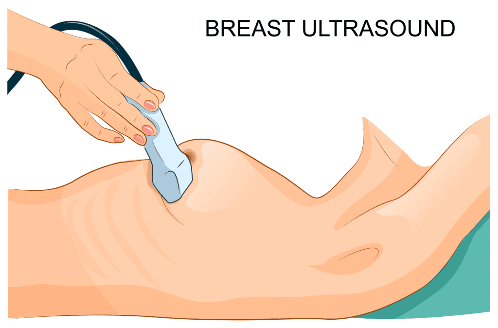
A Start-To-Finish Guide To Performing A Breast Ultrasound
- blog
- Posted on
-
 by
Michelle Macauley
by
Michelle Macauley - 1 comment
If you’re new to Breast Ultrasound it can feel overwhelming, but it doesn’t have to be! This step-by-step Breast Ultrasound roadmap will help you conquer this exam, and ensure that you don’t miss anything important along the way.
Have you ever faced an Ultrasound exam that you’ve never done before and felt completely lost? As you stare at the unfamiliar anatomy on the image, you find yourself struggling to remember back to that elusive lecture back in your school days. I’ve been there! I learned Breast Ultrasound the hard way, on the job, with archaic textbooks and through lots of trial and many missteps along the way.
Let me simplify that pathway for you! Journey with me as we dive into the technique of performing a Breast Ultrasound from start to finish.
Step 1: The Big Why
It all begins with a patient and a question to be answered. Why is the patient there and what is the reason for the exam? In breast imaging, there’s 3 big whys behind the order for an exam: screening for disease, addressing a patient symptom, or following up another test or previous study.
Patient Symptoms
Let’s start with patient symptoms. Is the patient experiencing breast pain, a palpable lump, nipple discharge, skin changes, nipple changes, axillary symptoms, issues with a breast implant, or have they experienced trauma to the chest?
Breast pain can present in 2 ways: focal or diffuse. Diffuse breast pain is felt throughout one or both breasts, and focal pain is pain that is felt within a specific area of the breast. Pain may or may not correspond to a woman’s monthly cycle, and can be either constant or intermittent.
A palpable lump is something that is felt with with the fingers. A lump may be a mass, a normal structure, such as a lymph node, or even normal breast tissue. A common misconception is that a lump represents a growth in the tissue, but by far, the most common finding when a patient presents with a lump is a mound of normal breast tissue.
Nipple discharge can be spontaneous, in which the discharge comes out by itself, or expressed, where the discharge needs to manually expressed. The most worrisome type of discharge is clear or bloody in color and spontaneous, though nipple discharge can commonly be associated with benign reasons, such as a papilloma.
Potential skin changes that can occur in the breast are redness, flaking, a rash, dimpling, ulcerations, swelling or peau d’orange appearance (skin pitting). Skin changes can be due to breast infection, or more sinister reasons such as inflammatory breast cancer. Nipple changes can include redness, flaking, crustiness, itching, or a rash. With nipple symptoms, a rare nipple cancer known as Paget’s disease must be ruled out.
Axillary symptoms may present as pain, a palpable lump, swelling or skin changes to the axilla. Common axillary findings include lymph node pathology, a skin mass, accessory breast tissue and/or a mass within the Axillary Tail of Spence. A normal lymph node can also present as a palpable Axillary lump.
An exam to evaluate implant integrity is ordered to evaluate a change in a breast implant such as breast hardness, swelling, redness, pain, or a change in breast contour. MRI is the gold standard test for evaluating the breast implant itself, but Mammogram and Ultrasound are commonly used to evaluate symptoms in a patient with breast implants, such as a palpable lump.
The last category of patient symptoms are related to breast trauma, either from an injury to the chest or from a breast procedure or surgery. After experiencing trauma, a patient can present with bruising, pain, or a palpable lump. Trauma, such a pocket of blood in the breast can also happen when patients are taking anticoagulants, which make them more susceptible to bruising.
Screening for Breast Disease
The gold standard test to screen for breast cancer is a Mammogram. Screening is performed when there’s not another imaging test or previous study being followed and in the absence of patient symptoms. A screening breast Ultrasound may be performed in patient’s with dense breast tissue, though this remains a controversial topic, or in patient’s that cannot undergo a Mammogram, such as a patient with a mental or physical disability that limits a patient’s mobility or tolerance for the exam.
Following-Up Another Imaging Test or Previous Study
If another imaging test, such as an MRI, CT or Nuclear Medicine study reveals a breast finding, then a Diagnostic Mammogram, often in conjunction with a Diagnostic Ultrasound, will be performed. If the patient is on a follow-up pathway, such a serial 6 month follow-up exams, then a Diagnostic Mammogram, Ultrasound, or both will be ordered, depending on the prior recommendations.
Other Relevant Patient History to Collect
Other essential patient history information to collect includes whether or not the patient has a personal or family history of breast, ovarian, or prostate cancer; any previous breast procedures including biopsies or surgeries; the patient’s age and menstrual status, including last menstrual period; whether the patient is currently pregnant, breastfeeding, or postmenopausal; the age that the patient first began menstruating; the age that the patient gave birth to their first child, when relevant; and whether or not the patient is taking any type of hormones, such as hormone replacement therapy, birth control pills, or has an IUD (especially the hormone-based type of IUD’s).
Once the reason for exam is ascertained, an a thorough patient history is collected, this information should be placed on a patient history worksheet. It’s then time to prepare the patient for the exam.
Step 2: Patient Positioning
The goals of proper patient positioning are to:
- Spread the breast tissue evenly over the chest wall
- Maintain consistent image optimization
- Achieve good contact of the transducer with the skin
- Decrease the depth needed to penetrate through the tissue (thin the breast tissue)
- Help prevent skin folds
- Create accurate and reproducible breast mass locations
- Ensure that the entire quadrant is evaluated (no tissue is missed)
Wedge Pillow
A wedge pillow is placed behind the patient’s back in order to support the patient’s back; accurately position the patient; help the patient stay as stationary as possible during the exam, and to ensure that patient positions are reproducible between different imaging facilities.
Right or Left Posterior Oblique
With a right or left posterior oblique position, the patient rolls slightly onto their side, and the patient’s ipsilateral arm (the arm corresponding to the side being scanned) is raised above the patient’s head. A wedge pillow is placed behind the patient’s back. This is the most common patient position used in Breast Ultrasound. The right or left posterior oblique position is used to evaluate the lateral breast (UOQ and LOQ), a breast that is medium to large in size, and the axilla. When the breast size is large, the right or left posterior oblique position is used to roll the breast away from the axilla.
Right or Left Lateral Decubitus
When using a right or left lateral decubitus patient position, the patient is rolled steeply onto their side and a wedge pillow is placed behind the patient’s back. The ipsilateral arm is placed above the patient’s head. The right or left lateral decubitus position is used to evaluate the lateral breast (UOQ and LOQ) and the Axilla when the breast size is very large. It’s too steep of an angle for the majority of breast imaging.
Supine
For a supine patient position, the patient lies flat on their back, with the ipsilateral arm raised above the patient’s head. The supine patient position is used to evaluate the make breast, a breast that is small in size, the medial breast (UIQ and LIQ), and the axilla (when the breast size is small to medium and the breast does not need to be rolled out of the way of the axilla).
Upright
With an upright position, the patient is placed in a sitting-up or a semi-erect position. The arms remain down by the patient’s sides. Note: this position thickens the breast tissue, causing uneven penetration throughout the breast and should only be used when necessary. An upright or semi-erect position is used to evaluate the superior breast (UOQ and UIQ) when the breast size is large, and to interrogate tissue that’s located above the breast (far superiorly), such as when there’s a palpable lump in this area.
Step 3: Prepping the Ultrasound Machine
Start by entering the patient’s information into the Ultrasound machine, including the patient’s name, date of birth and medical record number. Some machines auto-populate this information when the patient’s name is selected from a list. Next, select the proper transducer. For Breast Ultrasound, a high frequency linear transducer is used, usually with a range of 12-16 MHz.
The next step in setting up the machine is to select the correct exam type, known as a preset. The Breast Ultrasound preset can be found located under the small parts category. Image and transducer orientation is the next thing to consider. Each Ultrasound screen has a marker that delineates the active side of the FOV (field of view), or image. This icon should match the orientation of the active side (notch) of the transducer.
Step 4: The Structures and Layers of the Breast on an Ultrasound
Now let’s explore what in the world you are looking at on the Ultrasound screen. The structures and layers on a Breast Ultrasound image (from anterior to posterior) are:
Skin Layer
The skin layer is the most anterior layer on the Ultrasound image and it’s a thin layer, measuring less than 2 mm in thickness when normal. It’s hyperechoic in echogenicity compared to the medium gray color of the fat in the breast
Subcutaneous Fat Layer
The breast tissue is sandwiched between two fat layers: the subcutaneous fat layer and the retroglandular fat layer. When the breast density level is low and the tissue is more fatty in composition, then the three breast tissue layers (subcutaneous fat, glandular tissue and retroglandular fat) can be challenging to distinguish as separate, distinct layers. The subcutaneous fat layer is located below the skin layer and should be medium gray in echogenicity on the Ultrasound when the image is properly optimized.
Cooper’s Ligaments
Cooper’s ligaments are the support network of the breast, running from fascial plane to fascial plane in the breast throughout the 3 breast tissue layers. They are highly visible on the Ultrasound as hyperechoic lines running through the fat layers. Cooper’s ligaments also run through the glandular tissue layer, but are not visible as the glandular tissue is the same echogenicity as the cooper’s ligaments.
Glandular Tissue Layer
The glandular tissue layer, also known as the mammary layer lies posterior to the subcutaneous fat layer. It is hyperechoic in color, and anechoic milk ducts can often be visualized within this layer as thin, linear tubes. Depending on the density level of the breast tissue, the glandular tissue may either be a distinct layer of white tissue, or it may consist only as scattered patches of hyperechoic tissue.
Retroglandular Fat/Space
The retroglandular fat layer is located posterior to the glandular tissue layer and it’s medium gray in echogenicity. When the breast tissue is dense, it compresses this layer and it’s considered a space rather than a distinct tissue layer. When visualized, it appears medium gray in echogenicity, though it may appear darker gray in color due to shadowing from the glandular tissue anterior to it.
Chest Wall Structures
The chest wall structures are located below the retroglandular fat/space and include the pectoralis muscles, ribs, intercostal muscles, the pleura and lungs.
Pectoralis Muscles
The pectoralis major and minor muscles are immediately posterior to the retroglandular fat. The pectoralis minor muscle only runs below a small percentage of the breast, and is visible when scanning the UOQ of the breast. The pectoralis major muscle is visible throughout the entire breast. The pectoralis major muscle is anterior to the pectoralis minor muscle. On the Ultrasound, muscle is striated (linear white muscle fibers) in appearance.
Ribs
Ribs are the next chest wall structure and are located posterior to the pectoralis muscles. Strong posterior shadowing can be visualized below the ribs. The anterior surface of the rib will display a bright echogenic line, delineating a sharp interface in tissue type between a rib and the pectoralis muscle above it. This hyperechoic line can often be mistaken for the pleura.
Intercostal Muscles
The intercostal muscles run between the ribs and are striated in appearance on the Ultrasound. They intercostal muscles are located below the pectoralis major and minor muscles.
Pleura
Located below the ribs and intercostal muscles is the pleura, the bright, echogenic lining of the lungs. The pleura can be mistaken for the echogenic tissue interface that appears anterior to bone (ribs) on an Ultrasound. To distinguish between the two, look to see what appears below the white line: if it’s a dark area with strong posterior shadowing, then this is rib and the white line is the anterior surface of a rib; if it’s a gray, hazy area, then this is lung, and the white line is the pleura, which is the lining of the lungs.
Lung
Posterior to the pleura is the lungs, which appear as a hazy, gray shadow with air artifact on the Ultrasound image. Air artifact looks like an area of gray shadowing interspaced with hyperechoic areas. It’s distinctly different (brighter) than the dark gray shadowing that’s located posterior to ribs.
Step 5: Optimizing a Breast Ultrasound Image
The next step to performing a Breast Ultrasound is optimizing the image, in order to produce high-quality diagnostic images. Adjust the following Ultrasound controls to obtain the perfect image every time:
- FREQUENCY: adjust so that the highest frequency that allows penetration down to the chest wall is used
- GAIN: adjust so that the fat in the breast is a medium gray color
- DEPTH: adjust so that the pectoralis muscle is located about 2/3 of the way down the Ultrasound image
- OUTPUT POWER: adjust so that the lowest output power that produces a high quality diagnostic image is used
- TGC: adjust so that there’s equal brightness at all depths, and that each tissue type retains its unique echogenicity level (i.e. fat is medium gray, glandular tissue is hyperechoic)
- FOCAL ZONES: multiple focal zones (3-4) should be used for high image resolution throughout the image, with the lowest focal zone placed at the level of the pectoralis muscle
- TRANSDUCER PRESSURE: transducer pressure should be firm enough to eliminate cooper’s ligament shadowing and to produce a crisp Ultrasound image.
For more in-depth information on optimizing a Breast Ultrasound, check out the following post: HOW TO OPTIMIZE A BREAST ULTRASOUND IMAGE: 7 PITFALLS
Need a knobology refresher? Read this post: ESSENTIAL ULTRASOUND CONTROLS THAT EVERY SONOGRAPHER SHOULD KNOW AND HOW TO USE THEM: PART 1
Step 6: Annotating a Breast Ultrasound Image
Annotation refers to the words that are placed on an Ultrasound image to delineate the area of interest (organ), the location within the structure and the slice that is being taken through the structure by the transducer (transducer orientation). The most important thing about annotation for a Sonographer is to describe, not diagnose. For example, if multiple masses were located within the same quadrant of the breast, they can be labeled as mass 1, mass 2, and so on, to differentiate between them, rather than fibroadenoma 1, fibroadenoma 2, etc. Use the following list to annotate Breast Ultrasound images:
Location
The first step to annotating a Breast Ultrasound image is to list the location that’s being scanned, either the right or left breast or the axilla. This can be abbreviated: Rt Breast, Lt Breast or Rt Ax, Lt Ax.
Quadrant
The breast tissue can be divided into 4 quadrants. UOQ (upper outer quadrant), LOQ (lower outer quadrant), LIQ (lower inner quadrant), and UIQ (upper inner quadrant). This is most commonly used to annotate normal images, a set of images taken to document that an area of the breast was interrogated and that no pathology was visualized within that quadrant. Either quadrant or clock position should be used to annotate an image, not both.
Clock Position
Standard Breast Ultrasound annotation includes a clock position, where the breast is labeled like a clock, with 12:00 at the top of the breast, 6:00 at the bottom of the breast, and 3:00 and 9:00 representing the medial and lateral aspects of the breast. Only whole and half hours are used, no quarter hours
Distance from Nipple
Distance from the nipple can be annotated using one of two methods: centimeters from nipple or the ABC/123 method.
Centimeters From Nipple (CM FN)
The centimeters from nipple method delineates an exact distance from the nipple. The linear transducer surface is used as a ruler (most linear transducers are 5 cm across) to measure how far away a location or mass is from the nipple in centimeters. A mass located 8 centimeters from the nipple could be annotated: 8 CM FN, 8 + N (n=nipple), or 8N.
ABC/123 Methods
The ABC and 123 methods of labeling delineate a zone of tissue that specifies either the distance from the nipple or the depth in the tissue. These are more archaic methods of labeling, since they do not provide an exact location, but only a zone of tissue. They are rarely in use today, primarily being replaced by the centimeters from nipple designation.
- 123 METHOD: with the 123 method, concentric circles are drawn around the breast. Circle number one represents the zone of breast tissue closest to the nipple; circle three represents the zone of tissue around the periphery of the breast; and circle two represents the tissue is the middle zone of the breast, between circles one and three.
- ABC METHOD: with the ABC method, the breast tissue is divided into 3 depths. The A zone represents the tissue that’s located most superficially in the breast, B is mid depth, and C is the deep tissues of the breast.
To label using the ABC/123 methods, one number and one letter are combined to designate an approximate location. For example, 3B would represent a location around the outer periphery of the breast at mid depth in the tissue. The ABC and 123 methods are always used in conjunction with each other. The final annotation would read something like this: Rt Breast, 2:00, 3B, RAD, to delineate a mass in the Right Breast at the 2:00 position, around the outer periphery of the breast at mid depth in the tissue. You can see how this gives a much less precise location than Rt Breast, 2:00, 8 CM FN, RAD.
Transducer Orientation
The last piece of information that should be listed when annotating an Ultrasound image is the transducer orientation (the slice of information that’s being taken through the tissue, or to think of it in different terms, the direction is which the Ultrasound transducer is facing. There’s 4 transducer orientations used in Breast Ultrasound:
Sagittal (SAG):
The sagittal transducer plane takes vertical slices throughout the breast tissue and it is used when taking normal images behind the nipple and while scanning the axilla.
Transverse (TRV):
The transverse plane takes horizontal slices throughout the breast tissue (horizon= flat, side-to-side). It is used as a plane of imaging that’s perpendicular to the sagittal plane and most commonly used in the breast as the perpendicular plane to sagittal slices, for documenting normal subareolar images and when evaluating the axilla.
Radial (RAD):
The radial plane is the most important plane in breast imaging because it follows the course of the milk ducts, where most suspicious breast pathology occurs. The radial transducer plane extends in lines from the nipple to the periphery, like rays of the sun or spokes on a wagon wheel. It is the most accurate slice of information through the breast tissue because it gives a true representation of mass size and shape as it parallels the lie of the milk ducts.
Anti-Radial (ARAD):
The anti-radial imaging plane is the orientation that’s 90 degrees orthogonal or perpendicular to the radial plane. Radial and anti-radial planes are transducer orientations that are unique to the breast, and used solely in Breast Ultrasound.
Transducer Orientation Tips & Tricks
A few caveats to mention when using different transducer orientations in the breast:
- It’s essential to use transducer planes that are perpendicular to each other (sagittal and transverse, or radial and anti-radial). If transducer planes that are not perpendicular to each other are used, then the true length, height and width of a mass will not be demonstrated.
- When sagittal and transverse planes are used in Breast Ultrasound, mass size and shape can be under-represented since these transducer planes do not follow the lie of the milk ducts.
- Sagittal and transverse planes in Breast Ultrasound are primarily used to image the axilla, and for documenting normal images in the subareolar region.
Looking for a tutorial on scanning the breast in different transducer planes? Here’s a helpful guide: TRANSDUCER ORIENTATION AND BREAST ULTRASOUND: STOP DOING THIS
Step 7: Breast Ultrasound Protocols
It’s important when scanning an Ultrasound to have a set of protocols for documenting a normal exam and any pathology encountered along the way. Let’s explore 3 scenarios and which images should be taken during each exam.
Screening Breast Ultrasound
A screening Breast Ultrasound is performed in the absence of symptoms and when there is no need for a follow-up exam to screen for the presence of disease. The breast is split into 4 quadrants: UOQ, UIQ, LIQ, LOQ. Each quadrant is scanned independently in both the radial and anti-radial planes. If pathology is encountered, it should be documented and measured in both the radial and anti-radial planes, with and without color Doppler images.
A negative image in both transducer planes is taken after pathology documentation (and also when there is no pathology visualized) to indicate that the entire quadrant was evaluated. The process is repeated for each quadrant of the breast, and in the opposite breast. An Ultrasound of the axilla may or may not be performed during a screening breast Ultrasound, depending upon the protocols of the clinical site.
Targeted Breast Ultrasound
A targeted Breast Ultrasound is answering a specific question. It is used to evaluate patient symptoms, to follow-up a Diagnostic Mammogram or other test, such as an MRI, or as a means of conducting serial follow-up exams (such as a 6 month follow-up protocol).
When performing a targeted Breast Ultrasound, only the quadrant or quadrants that the contain the AOI (area of interest) are evaluated. The area of interest is the follow-up site, the area in question on a Mammogram, MRI or other test, or the area of the patient’s symptoms. First the AOI is evaluated in both the radial and anti-radial planes, and when pathology is visualized it is imaged, measured and the vascularity pattern is assessed in both planes with color Doppler.
Next, the quadrant that the AOI is in should be evaluated, and any additional pathology should be documented. Finally, a negative image denoting the quadrant being interrogated should be documented in both the radial and anti-radial planes, attesting that the entire quadrant was thoroughly evaluated.
Axillary Ultrasound
An Axillary Ultrasound is performed either as part of a screening Breast Ultrasound, or to follow-up findings visualized on a Mammogram or other imaging test, as part of a 6 month follow-up protocol, or to evaluate patient symptoms. The entire axilla should be scanned in the sagittal and transverse planes, ensuring that the Axillary Tail of Spence, which is the normal extension of glandular tissue into the axilla, is included within the scan.
It is essential to visualize the axillary lymph nodes and to characterize their appearance. Normal axillary lymph nodes have all of the following features on an Ultrasound:
- Thin outer cortex (<3-4 mm in thickness)
- Fatty, central hyperechoic hilum
- Hilar blood flow pattern (blood flow towards the central portion of the lymph node)
Abnormal lymph nodes can display any of the following features on an Ultrasound:
- Thickened outer cortex (>3-4 mm in the A-P, or height dimension)
- Outer cortex with irregular margins (angular or microlobulated)
- Rounded shape
- Loss of the central fatty hilum
- Peripheral vascularity (flow in the cortex of the node vs the hilum)
- Focal cortical bulge (macrolobulated margin)
If all lymph nodes appear normal, one normal lymph node should be documented and measured in both the sagittal and transverse planes with and without color Doppler images. If abnormal lymph nodes are visualized, then the 3 most abnormal lymph nodes should be documented, measured and evaluated with color Doppler in both transducer orientations.
If Accessory Breast Tissue corresponds to a palpable area, then the Accessory Breast Tissue should be documented in the same manner that pathology is imaged: in two planes, measuring the size of the patch of Accessory Breast Tissue, and evaluating it with and without color Doppler in both orientations. Special care should be taken to evaluate the Accessory Breast Tissue for the presence of a mass within the patch of tissue.
If pathology is evaluated in the axilla, such as a mass in the skin, in the Axillary Tail of Spence, or within a patch of Accessory Breast Tissue, then the mass should be imaged, measured and evaluated with color Doppler in two planes. After lymph node and pathology documentation, two normal images should be taken of the axilla in both the sagittal and transverse planes to demonstrate that the entire axilla was evaluated.
Need a crash course on characterizing lymph nodes? BREAST ULTRASOUND: THE BEGINNER’S GUIDE TO FINDING AND CHARACTERIZING LYMPH NODES
Step 8: Characterizing Breast Masses
Rule number 1 when characterizing a breast mass is that not all masses follow the “rules,” meaning that Ultrasound features of benign vs malignant disease are generally true, but not always accurate. It’s important to watch out for the wolf in sheep’s clothing- a breast mass that appears benign at first glance, but that is really malignant in nature.
When characterizing a breast mass, the most suspicious feature of a mass is used to categorize the overall suspicion level of the mass. Characterizing breast masses on an Ultrasound is a crucial step when scanning, as it helps determine if there’s any additional areas that need to be evaluated. The following criteria can be used to characterize the Ultrasound features of a mass:
Cystic vs Solid vs Complex
- CYSTIC: a cystic mass is a fluid-filled mass. It’s known as a simple cyst when it’s anechoic on the Ultrasound, indicating that the cyst is filled with a clear, watery substance. A cyst may also be known as a complicated cyst when it has debris within the cyst, which can be mobile or non-mobile in nature. Internal debris may have a fluid/debris level, display low-level gray echoes throughout the cyst which can mimic the appearance of a solid mass, or have floating, mobile internal echoes inside. A cystic mass can be filled with thin, watery fluid, serous fluid, blood or pus, and/or be a blood vessel.
- SOLID: a solid mass is composed of solid tissue rather than fluid. On Ultrasound, a solid mass will be hypoechoic, with dark gray echoes inside the mass. A solid mass can be either benign or malignant.
- COMPLEX: a complex mass has both solid and cystic components on the Ultrasound. It will be heterogeneous in echotexture, with an uneven pattern of internal echoes and multiple echogenicities displayed on the Ultrasound.
Breast Tissue Planes & Mass Orientation
Breast Tissue Planes
The tissue planes in the breast run horizontally on the Ultrasound image, parallel to the skin and the chest wall layers. Tissue planes are important in Breast Ultrasound because benign breast masses respect the tissue planes, while malignant masses do not.
Mass Orientation
There are 2 mass orientations on a Breast Ultrasound: horizontal and vertical orientation. Benign breast masses displace, or push tissue planes planes out of the way. Malignant breast masses invade adjacent tissue planes, crossing tissue planes and fixing the surrounding tissues to them.
- HORIZONTAL ORIENTATION: a breast mass that has a height (A-P, or anterior-posterior dimension) that’s smaller than its length is demonstrating that it’s respecting the tissue planes by displacing them. This is known as a horizontal or wider-than-tall orientation.
- VERTICAL ORIENTATION: a breast mass that has a height (A-P dimension) that’s larger than its length is demonstrating that it’s no longer respecting the tissue planes by invading the adjacent planes. This is known as a vertical or taller-than-wide orientation.
Breast Mass Shape, Margins & Borders
The shape of a breast mass is its form or substance. The margins of a mass are the edges of the mass itself, and the borders of a breast mass refers to the tissue that is immediately surrounding a mass.
Breast Mass Shape
There are 3 breast mass shapes: oval, round and irregular.
- OVAL: oval shapes have a horizontal orientation within the breast tissue, with a height that’s smaller than its length, and are most commonly benign.
- ROUND: round shapes can be either benign or malignant. A mass with a round shape has an A-P dimension that’s equal to its length, which may be an early indicator that a mass is growing in a vertical nature and starting to no longer respect the tissue planes. It’s important to carefully scrutinize round masses to ensure that they are not displaying any other worrisome Ultrasound features.
- IRREGULAR: irregular masses in breast imaging are shaped like an ink blot, demonstrating that they are no longer respecting the tissue planes, which is a suspicious mass feature on an Ultrasound.
Breast Mass Margins
There are multiple categories of breast mass margins, including circumscribed, non-circumscribed, macrolobulated, microlobulated, angular, and spiculated.
- CIRCUMSCRIBED: a circumscribed mass has even, well visualized margins, and is most commonly benign in nature. With a circumscribed mass, you can trace the outline of the mass in its entirety, with no hazy or obscured segments.
- NON-CIRCUMSCRIBED: a non-circumscribed breast mass has one or more areas along the margin that are hazy, ill-defined or obscured. This is most commonly associated with suspicious masses on Breast Ultrasound.
- LOBULATED: lobulations are rounded protrusions extending from a mass. The lobulations can be solitary or multiple, and can be small (<2mm) or large (>2mm).
- MACROLOBULATED: a mass with 3 or less large lobulations has macrolobulated (macro= large) margins. Macrolobulated masses are shaped like a cloud and are most commonly associated with benign breast masses.
- MICROLOBULATED: a mass with multiple, small lobulations has microlobulated margins. It is shaped like the petals of a carnation flower, and is usually associated with malignant breast masses.
- ANGULAR: a breast mass can also present with pointed margins, known as angular margins. Angular margins are shaped like the edges of a starfish, and are a malignant feature for a breast mass on Ultrasound.
- SPICULATED: a mass with long, linear extensions, known as spiculations, is known as a spiculated mass. Spiculations are the most suspicious feature of a breast mass, and the mass will be shaped like an octopus, with a body and leg-like extensions.
Breast Mass Borders
There are 4 categories of breast mass borders on a Breast Ultrasound: no/partial border, a pseudo-capsule, a true capsule and desmoplasia.
- PSEUDO-CAPSULE: the most common benign breast mass border is known as a pseudo-capsule, a thin, echogenic line around a breast mass on an Ultrasound. It is known as a pseudo-capsule because histologically, it does not form a complete capsule around a mass.
- TRUE CAPSULE: a mass with a true capsule also displays a thin, hyperechoic line around it on an Ultrasound image. It is indistinguishable from a pseudo-capsule except for histologically, where it forms a complete capsule around the mass. A lipoma is the only mass that has a true capsule on a Breast Ultrasound, which cannot be visualized when a lipoma is hyperechoic. So most of the masses that have a thin capsule on a Breast Ultrasound will be pseudo-capsules.
- NO/PARTIAL BORDER: a breast mass without a border, or with only a partial border, can be either benign or malignant. It should be carefully interrogated however, because breast masses first start expanding (via irregular margins) along their sides on an Ultrasound because it’s the path of least resistance. A mass can expand laterally without crossing tissue planes, while vertical growth requires invading adjacent tissue planes. This is why a horizontal orientation is associated with tissue displacement and a vertical orientation is associated with tissue invasion. A mass with no border, or with a border only along the anterior and posterior edges may be displaying the first signs of irregular growth along its medial or lateral edges. A mass with borders that are only visualized along the anterior and posterior edges may also be benign. Axial resolution on an Ultrasound is superior to lateral resolution, resulting in clearer visualization of the edges of a mass along the anterior and posterior edges vs the sides of a mass. So non-visualized borders along the sides of a mass may simply be due to the limitations of the Ultrasound itself- inferior lateral resolution- rather than the beginning stages of irregular margin formation.
- DESMOPLASIA: a malignant breast mass border is known as desmoplasia. Its Ultrasound appearance is a thick, echogenic halo around a breast mass with irregular, hazy, indistinct margins. The first mode of spread of a breast mass is by infiltrating adjacent tissues via microlobulated, spiculated and angular margins. The body in turn, launches a host response, in an effort to halt the expansion of the mass. During a host response, the tissue surrounding the mass becomes fibrotic. The combination of these irregular mass margins from the mass expanding, and the tissue fibrosis from the host response forms a thick, echogenic halo around a suspicious breast mass on an Ultrasound. Note that some masses grow rapidly, and are located before the body has had time to launch a host response. So not every malignant breast mass will display a desmoplastic reaction.
Did you know that desmoplasia should be included in the measurements of a breast mass, but spiculations should be excluded? For additional measuring tips and trick, check out this article: MEASURING BREAST MASSES ON ULTRASOUND: 10 MISTAKES TO AVOID
Breast Mass Echo Pattern
The echo pattern of a mass describes the brightness and the uniformity of the echoes inside a mass and the degree of attenuation associated with the mass. Breast mass echogenicity, echotexture and posterior echo patterns are considered weak indicators of benign vs malignant disease when used as a sole criterion, as they can be associated with both benign and malignant breast disease.
Breast Mass Echogenicity
There are 5 echogenicity categories used in Breast Ultrasound: anechoic, hyperechoic, hypoechoic, isoechoic and complex. A sound wave goes into the tissue, encounters a mass, and a portion of that wave returns back to the transducer. The degree of sound wave reflection determines the brightness of the echoes on the Ultrasound image, which is the color of the mass, or its echogenicity. The echogenicity, or brightness of a structure is determined by comparing the color of the structure to the medium gray echogenicity of the fat in the breast. Anechoic, hyperechoic and isoechoic are more common in benign masses, and hypoechoic and complex tend to be associated masses that are more suspicious in nature.
- ANECHOIC: an anechoic mass is black in color on the Ultrasound, indicating an absence of returning echoes from the tissue.
- HYPERECHOIC: hyperechoic masses are white to light gray on an Ultrasound, indicating a strong reflection of the sound waves.
- HYPOECHOIC: a hypoechoic mass is a weak reflector on an Ultrasound and appears as a dark gray color on Ultrasound. As a general rule, the more hypoechoic a mass on a Breast Ultrasound (known as markedly hypoechoic, or darker gray), the higher the suspicion level of the mass.
- ISOECHOIC: an isoechoic mass is medium gray in color, and has an echogenicity similar to the fat in the breast, indicating that a mass has a similar amount of echoes reflecting back to the transducer as the fat layer.
- COMPLEX: complex has 2 meanings: it indicates that a mass has multiple brightness levels present (multiple echogenicities); it also means that a mass has both solid and cystic components.
Breast Mass Echotexture
The echotexture of a mass refers to the uniformity of the brightness levels within a mass.
- HOMOGENEOUS: a benign echotexture is known as homogeneous, which indicates that a mass has a uniform pattern throughout the mass. The echoes within the mass are the same color throughout the mass.
- HETEROGENEOUS: a more worrisome type of echotexture is heterogeneous, in which a breast mass displays multiple levels of brightness on an Ultrasound, in an uneven pattern of echoes.
Breast Mass Posterior Echo Pattern
The echo pattern of a mass describes the degree of sound wave attenuation through a mass on an Ultrasound. Attenuation is the loss in the strength of a sound wave as it travels through a dense mass or deeper and deeper into the body.
- SHADOWING: shadowing presents as dark echoes posterior to a mass on an Ultrasound. It indicates increased attenuation of the sound waves through a dense mass. Shadowing can be found with both benign and malignant masses.
- ENHANCEMENT: enhancement on an Ultrasound appears as bright echoes posterior to a mass, almost as if a flashlight is being shined down through the mass. It indicates no attenuation of the sound waves as they travel through a mass, and most commonly occurs with masses that have some sort of cystic component. Enhancement can be found in both benign and malignant masses.
Breast Mass Vascularity
The vascularity of a mass is the amount and the pattern of blood flow within a mass. There are several categories of vascularity that a mass may display, including: avascularity, hypovascularity, hypervascularity, peripheral vascularity, central vascularity and internal vascularity.
- AVASCULARITY: avascular means that a mass has no vascularity. It is the absence of vascularity within a breast mass, which presents as no color or power Doppler signal within the Doppler box. Avascularity can be associated with both cystic and solid masses. It’s important to note that the absence of vascularity does not confirm that a mass is cystic. Avascularity is most commonly associated with benign breast masses.
- HYPOVASCULARITY: a mass that is hypovascular displays a weak vascularity pattern on an Ultrasound. It is a small amount of color or power Doppler signal displayed within a breast mass. It is most commonly found with benign breast masses.
- HYPERVASCULARITY: hypervascularity is a large amount of blood flow within a mass on a Breast Ultrasound, and is displayed as a large amount of color or power Doppler signal within a Doppler box. Hypervascularity can be found in malignant masses, ductal masses, and with infection and inflammation in the breast. The concern with hypervascular masses, is neoangiogenesis, the concept that malignant masses grow their own blood supply to fuel their rapid growth.
- INTERNAL VASCULARITY: internal vascularity means that a mass has vascularity inside the mass on a Breast Ultrasound. A mass that has internal vascularity confirms that the mass is solid tissue, and may be either benign or malignant in nature. A mass with internal vascularity does increase the scrutiny level of the mass, however, and the mass should be carefully evaluated for any additional suspicious Ultrasound features.
- PERIPHERAL VASCULARITY: with peripheral vascularity, small blood vessels taper around the edges of a mass. This is most commonly found with benign breast masses, and with suspicious lymph nodes.
- CENTRAL VASCULARITY: malignant breast masses and normal lymph nodes most commonly display central vascularity, which is the presence of large, feeding vessels oriented towards the central portion of a mass or lymph node.
Step 9: Breast Mass Measuring Basics
The most important aspects of measuring a breast mass are measuring the longest lie of the mass (longest length), even if it means the measurement is in a slightly oblique plane; ensuring that the measurements are perpendicular to each other in complementary scanning planes (SAG/TRV vs RAD/ARAD); and measuring a length (horizontal measurement), a height (vertical measurement) and a width (horizontal measurement).
The most common incorrect measurements involve measuring the height dimension twice (two vertical measurements instead of two horizontal measurements and only one vertical measurement) which means that not all mass dimensions have been represented; calipers that are not placed perpendicular to each other, which also means that mass dimensions are not accurately portrayed; transducer planes that are not perpendicular to each other being used, which means that non-complementary slices of a mass are being taken; and shortening the dimensions of a mass by not measuring the longest lie, or length of a mass.
For more breast measuring tips and tricks, check out this guide: MEASURING BREAST MASSES ON ULTRASOUND: 10 MISTAKES TO AVOID
Step 10: You’ve Found a Breast Mass. Now What?
When a mass is visualized on a Breast Ultrasound, it’s important to consider the relationship of the mass to the surrounding structures and layers and when a mass has suspicious features, to also to evaluate the axillary lymph nodes for the presence or absence of metastatic spread to the nodes. Here’s a list of what to evaluate next when you encounter a breast mass with suspicious features on an Ultrasound:
Nipple Invasion
It’s important to demonstrate the relationship of a breast mass to the nipple. Is the mass connected to the nipple? Is the mass close to, but not visibly connected to the nipple? Is the mass contiguous with the nipple, meaning that it’s hard to differentiate where one begins and the other ends?
If a mass is not clearly connected to but is located close to the nipple, then a measurement should be taken delineating the distance between the mass and the nipple on the Ultrasound. The nipple itself should also be clearly labeled on the Ultrasound images to demonstrate its position relative to the mass.
Chest Wall Invasion
It’s also critical to demonstrate the relationship of the mass to the chest wall structures (the pectoralis major muscle is used as a landmark). Is the mass fixed to the pectoral fascia? Is the mass close to, but not fixed to the chest wall?
Not sure? Have the patient take in a deep breath and then release it. Watch for movement of the mass independent of the muscle. If the mass is close to but not fixed to the chest wall, the mass will move independently of the muscle. If the mass is not fixed to the pectoral fascia, then measure the distance between the mass and the pectoralis major muscle to demonstrate their relationship to one another.
Skin Invasion
Demonstrating the relationship between a mass and the skin layer is also a critical piece of information in Breast Ultrasound. This helps reveal whether a mass is a skin mass vs a breast mass.
A superficial mass on a Breast Ultrasound may be completely contained within the skin layer (considered a skin mass); partially within the skin layer and the subcutaneous fat layer (considered a skin mass); located just below the skin layer but communicating with the skin layer via a track to the skin (considered a skin mass); or located completely within the subcutaneous fat layer with not communication to the skin layer (considered a breast mass).
When a breast cancer spreads to the skin layer, the skin layer thickens, and multiple hypoechoic linear extensions can be visualized connecting the mass to the skin layer. The skin layer can also be thickened on the Ultrasound when a benign sebaceous cyst, which is a skin mass, is inflamed or infected.
When a superficial breast mass is visualized, it’s essential to decrease the depth and take close-up images of the the relationship between the mass and the skin layer, and color Doppler images looking for the presence of internal vascularity. Though internal vascularity can be visualized in both a suspicious superficial mass and an inflamed skin lesion.
Relationship to the Milk Ducts
Whether or not a mass is ductal in nature is an important feature of a breast mass. When a breast mass is visualized anywhere in the breast tissue, but especially when near the nipple in location, the ends of the mass, in a radial plane should be carefully interrogated to determine whether there’s a segment of milk duct extending from either end of the mass. Any visible ductal segments should be excluded from the measurements of the mass.
It’s also important to note that a hallmark feature of ductal masses, both benign and malignant is hypervascularity. The presence or absence of calcifications (white dots) within the ductal mass should also be noted.
If a suspicious mass is ductal in nature (has a ductal segment extending from one or both ends of the mass), then it is also important to delineate the direction of breast mass growth, either branch pattern or duct extension.
Branch pattern indicates that a ductal mass is spreading away from the nipple. The blind end of the mass (rounded edge of the mass) will be on the side towards the nipple, while the branching end of the mass (angular or spiculated margins) will be located away from the nipple.
Duct extension is the growth of a ductal mass towards the nipple. The blind end (rounded edge) of the mass will be located away from the nipple, while the branching end of the mass will be branching towards the nipple.
Satellite Nodules
After visualizing a suspicious breast lesion and determining its relationship to nearby structures, the next thing that should be evaluated is the presence or absence of any satellite nodules. A malignant breast mass first infiltrates adjacent tissues and structures via irregular margins, and next forms separate growths of variable distance from the primary mass.
At a minimum, the quadrant that the primary mass is located in should be evaluated for the presence of satellite masses. Any satellite masses should be imaged and measured in two planes and evaluated with color or power Doppler. The distance between the satellite mass and the primary mass should be measured. If the primary mass is located near the border of two quadrants, such as near 12:00, then both adjacent quadrants should be evaluated for the presence of satellite nodules.
Axillary Lymph Node Evaluation
Whenever a suspicious breast mass is located on an Ultrasound, an important step is to evaluate the axillary lymph nodes, looking for any signs of metastatic spread to the nodes.
Normal lymph nodes have a thin, hypoechoic outer cortex (less than 3-4 mm in thickness), a central hyperechoic fatty hilum and a hilar blood flow pattern
Abnormal lymph nodes can display a thickened outer cortex, rounded shape, irregular cortical margins, loss of the central fatty hilum, a focal cortical bulge, and/or blood flow around the periphery (cortex) of the lymph node.
Learn the ins and outs of Breast Ultrasound lymph node characterization through this tutorial: BREAST ULTRASOUND: THE BEGINNER’S GUIDE TO FINDING AND CHARACTERIZING LYMPH NODES
Step 11: Completing the Sonographer Worksheet
Completing the Sonographer worksheet accurately, in a logical and succinct manner, and knowing what to include and what not to include on the worksheet is essential to ensure good communication with the Radiologist. In Breast Imaging, the Radiologists are primarily on-site and Breast Ultrasound exams are most commonly presented in-person to the Radiologists, though this is not the case at every site.
Regardless of whether an exam is being presented in-person or sent off-site to be read by the Radiologist later, a complete and accurate Sonographer worksheet ensures that vital information is communicated to the Radiologist (such as if you forget to present an important item), and that the Radiologist has a handy reference sheet to refer back to if they dictate the findings at a later point in time. The following categories highlight what to include, and what not to include on a Sonographer worksheet:
Reason For Exam
The most important part of a Sonographer worksheet is the reason for exam. Let me repeat that! The most important part of a Sonographer worksheet is the reason for exam! When no reason for an exam is given, and if that is accidentally carried over into a Radiologist’s report, a facility may risk not being reimbursed for that exam. That’s why we started this Breast Ultrasound journey with the big why. Why is the patient there and are they having any symptoms? This should go on your worksheet before you even begin scanning as this will help you know what to focus on during the exam.
Don’t Diagnose, Characterize
A Sonographer is not a doctor. It is not our job to diagnose. Do we know what we are looking at? Most of the time. Do we diagnose? No. Our job is to characterize.
When we find a mass in the breast, we should not write “fibroadenoma found in the right breast, 2:00, 5 CM FN.” Rather, describe what you found…”hypoechoic mass with internal vascularity and circumscribed margins found in the right breast at 2:00, 5 CM FN.”
This also goes for image annotation. When measuring a breast mass, do not label an image with “fibroadenoma” or another type of pathology. If you need to add a descriptor to delineate between different pathologies for measurement purposes, then use mass 1, mass 2, and so on.
The exception to this rule is when dealing with breast cysts. It’s ok to write simple cyst, complicated cyst or complex mass on a Sonographer worksheet (never on an Ultrasound image though) when describing the features of a breast cyst. We are not diagnosing in these instances, we are describing the category of the cystic area that we visualized, which the Radiologist can change within their report if they disagree with the category of the cyst.
Less Is More
A Sonographer worksheet is not a college dissertation, and this is not the time for a lengthy treatise. Focus on the important highlights. Ever hear the expression “a picture is worth a 1000 words?” Well your Ultrasound study is full of pictures! Let them speak for themselves and point out only the major findings.
This also applies to measuring. Don’t measure 10 breast masses in each breast, just don’t! Most commonly, the 3 most suspicious masses in each breast are measured and recorded on the Sonography worksheet. When deciding what to measure, consider the features- a large 4 cm simple cyst, a small mass with angular margins and a medium-sized mass with benign features can each tell the story of the pathology present within a breast better than measuring 5 small simple cysts and ignoring more suspicious and conspicuous findings.
Focus on What’s Abnormal, Not Normal
Please, please, please do not list every normal thing and structure that you visualized on your scan. For example, normal tissue visualized within the UOQ of the breast, and no pathology visualized within the milk ducts, skin layer and chest wall structures in the UOQ.
This goes back to the adage that a picture is worth a 1000 words. Your normal Ultrasound images will speak to all of the normal things that you encountered along the way. The only time that I write normal things on my Sonographer worksheet is when a document a single, normal axillary lymph node when evaluating the axilla. Otherwise, the focus should be on the abnormal, and describing those abnormal findings in clear, precise descriptions that are short, sweet and right to the point.
Don’t Forget Those Lab Values
There’s not many times that lab values come into play on a Breast Ultrasound Sonographer worksheet, unless there’s a patient with an infection, in which case a WBC count might be helpful information, or the type of pathogen that grew within a culture when dealing with an ongoing breast abscess. Lab values however, are critical for other types of Ultrasound exams, such as abdomen, GYN and OB. When relevant, don’t forget to include lab values on the Sonographer worksheet.
Tying It All Together
The last step in completing a Sonographer worksheet is a review. Did you answer the question that brought the patient in for the exam in the first place? For example, what did the patient’s lump correspond to (normal tissue, a breast mass, etc.). Many times, as a Sonographer, the exam will be normal, which is also an answer to the patient’s reason for exam, in a way, as it’s ruling out certain pathologies. Don’t forget to do a quick review before you turn in your Sonographer worksheet into the Radiologist to ensure that you have included all relevant information that answers the question of why that patient was there in the first place.
Conclusion
Successfully performing a Breast Ultrasound is much more than waving an Ultrasound wand over a patient’s skin, as any Sonographer can attest to. We are highly trained and highly specialized professionals. Yet with all that knowledge, it’s not uncommon to encounter a type of Ultrasound exam that we’ve never scanned before. This guide will serve as a helpful reference, so that you know what to do and why when a Breast Ultrasound comes your way.
Today we learned the keys to performing a Breast Ultrasound exam, from beginning to end, all the way from patient and machine set-up, through scanning the breast tissue and breast mass characterization, to the final step of completing an accurate Sonographer worksheet. When you next encounter a Breast Ultrasound, you now have the tools to say “that was easy!” Admit it, you’re picturing the Staples red easy button. Well go ahead and push it, because you have conquered this exam.
Thanks so much for the kind words! Much appreciated!