- Your cart is empty
- Continue Shopping
Essential Ultrasound Controls And How To Use Them: Part 4
This is Part 4 of the 4 part Essential Ultrasound Controls Every Sonographer Should Know Series. See the overview here.
Time to continue along in our journey of learning the art of great Ultrasound knobology. When a challenging scan comes our way, knowing how to utilize the Ultrasound controls in front of us can make or break the quality of the images that we produce. Let’s Ultrasound…Essential Ultrasound Control 16: Harmonics
Harmonics is a control on the Ultrasound machine which is either on or off on some machines, and is tied to the Frequency control on other machines. Meaning that when Frequency is adjusted, the Harmonics setting is also affected.Harmonics Defined
Turning on Harmonics reduces noise (extraneous echoes) in the Ultrasound image, resulting in a crisper image with reduced image artifacts. Harmonics sends and receives the Ultrasound signals at two different frequencies. The sound waves are transmitted into the body at one frequency but received back to the transducer at double the frequency. Higher frequencies provide higher image resolution, but at the expense of penetration.Pros And Cons Of The Harmonics Control
Pros of the Harmonics Ultrasound control include: cleaner image with less Ultrasound artifacts (shadowing, etc.). A smoother image with less noise (artifactual echoes or speckle). Cons of the Harmonics Ultrasound control include: very deep or very superficial structures are not well visualized with harmonics. Penetration is lost (information in the far field of the image drops out).Optimal Harmonics Setting For An Ultrasound
Using harmonics is similar to using the frequency control in a way, and it’s integrated into this control on some Ultrasound systems. You have to weigh the need to penetrate through the tissue against the need for high resolution. High resolution doesn’t get you much if you can’t penetrate deeply enough to see what you are trying to image. Since the returning frequencies are double that of the transmitted signals, this Ultrasound control is best suited to imaging more superficial body structures (thyroid, breast, etc.) rather than deeper structures (liver, aorta, etc.). My best advice? Try it out! When you turn it on, can you see deeply enough to visualize the posterior borders of the structure you are imaging? If so, great! You have enough penetration and can reap the rewards of a high resolution image. If you can’t see the posterior margin of the structure, then you need the ability to penetrate more than you need high resolution. Harmonics gives you higher image resolution which can decrease image artifacts and improve delineation of small or subtle findings, but it decreases the ability to penetrate deeply or through dense tissues.Other Names For This Control
Various names for this control among different manufacturers include: Harmonics, harmonic imaging, CHI (coded harmonic imaging), THI (tissue harmonic imaging), H (and can also be paired with Frequency: HPen, HRes, HGen).Essential Ultrasound Control 17: Compound Imaging
Some Ultrasound controls help us to reduce image artifacts, and compound imaging is one of those controls. The drawback though is that sometimes the presence of artifacts helps provide vital clues about pathology in the body. Use of compound imaging should be tailored to each portion of the exam, weighing the desired outcome of the image.Compound Imaging Defined
Compound imaging is a control on an Ultrasound machine which combines 3 or more frames from different steering angles into a single frame. Compound imaging smooths the image, eliminating image artifacts. On some ultrasound machines, compound imaging is either off or on; with other machines, the lecel of compounding that the machine applies to the image can be set to high, mid or low.Optimal Compound Imaging Level For An Ultrasound
Generally, compound imaging is set at a low level for most types of imaging. When a mass is visualized, compound imaging can be turned off to ensure that important features of the mass are not missed. When set too high, important characterizing artifacts (such as shadowing and enhancement) and subtle findings can be eliminated (smoothed) out of the image. For Breast Ultrasound in particular, compound imaging is typically kept on a lower setting, or off, depending on the exam objectives, as Breast imaging relies upon the presence or absence of image artifacts to characterize breast masses.Other Names For This Control
Various names for this Ultrasound control among different manufacturers include: crossXbeam, SonoMB, iBeam, SonoCT, OmniBeam, spatial compounding, CRI, Xview, ApliPure (don’t confuse with ApliPure+ which is an SRI Ultrasound control).Essential Ultrasound Control 18: SRI
Our next Ultrasound control is how tissues and structures on an image get their texture. It makes a liver look like a liver. Let’s learn how to harness its power…SRI Defined
SRI, or speckle reduction imaging, is a control on an Ultrasound machine that increases or decreases the amount of speckle (noise) in an image. Speckle is extraneous echoes (dots) within an image that do not correspond to actual structures. It’s the bright pixelated dots within an image which gives tissue it’s textured look on an Ultraosund. The SRI control eliminates weak signals (speckle) and boosts, or brightens strong signals (true echoes).Optimal SRI Level For An Ultrasound
When SRI is set too high, the image becomes smooth, which can eliminate subtle findings. When SRI is set too low, the image becomes too speckled (grainy), which also can make the detection of subtle finding hard to visualize among the many extraneous echoes. SRI should generally be set to a medium level for most types of Ultrasound image. If the image appears too textured looking, then slightly increase SRI. If the Ultrasound image seems really smooth, and tissue doesn’t display any texturing, then slightly decrease the SRI level.Other Names For This Control
Various names for this Ultrasound control among different manufacturers include: SRI, XRES, uScan, TissuePure, SONO HD, iclear, SRI HD, Adaptive Speckle Reduction, SRF, Mview, SCI, Aplipure+, TeraVisionEssential Ultrasound Control 19: Dual Screen
Our next Ultrasound control, once mastered can be used as a short cut. A manner of taking less images while still providing the same amount of information. It’s also a great comparison tool. Meet the Dual Screen Ultrasound control…Dual Screen Defined
Dual screen is a control on an Ultrasound machine which allows an image to be split into 2 or 4 images. This is primarily used with the image split in two. Dual screen allows information to be displayed side by side within the same Ultrasound image, which is handy for displaying measurements and for comparison. Here’s a list of what you can show with the Dual Screen Ultrasound control:- View 2 areas side-by-side to compare and contrast them (such as the Right and Left Parotid Gland)
- Measure an organ or pathology in all 3 dimensions and display it on one screen
- Demonstrate an area with and without Color or Power Doppler
- Feature a mass with and without Elastography
- In vascular ultrasound, show veins with and without compression
- Show an appendix with and without compression and Color Doppler
- Demonstrate an area with and without the Valsalva maneuver applied (such as when imaging a hernia)
How to Use the Dual Screen Control
Dual screen consists of two buttons, side-by-side. One is labeled right (R) and the other is labeled left (L). To use dual screen, press either the right or left handed side key to turn it on, then toggle between the two keys to activate each side of the screen. Only one side of the screen can be active at a time. Once you have toggled to the active side of the screen, move your transducer to take the image you want. Next, activate the other side of the image and then move your transducer to take the image that you need. Once both sides have the image that you want, press the freeze and then the print/store key to save the image.Other Names For This Control
Various names for this Ultrasound control among different manufacturers: dual screen, R/L.Essential Ultrasound Control 20: Panoramic Imaging
What do you do when the area that you are trying to image doesn’t fit on the Ultrasound screen? First, you switch to a sector field of view, if you are using a linear transducer. If the area of interest still won’t fit, what then? That’s when we use a handy, dandy Ultrasound control known as panoramic imaging.Panoramic Imaging Defined
Panoramic imaging is a control on an Ultrasound machine which allows the user to move the transducer over a wide area, covering a much wider area of interest than would fit into a standard FOV (field of view), and mesh that data into a panoramic image. Think of multiple frames of data being stitched together side-by-side to create a new, larger image. This is used to image structures of pathology that’s too large to fit into a traditional linear or sector field of view.How To Use The Panoramic Imaging Control in Ultrasound
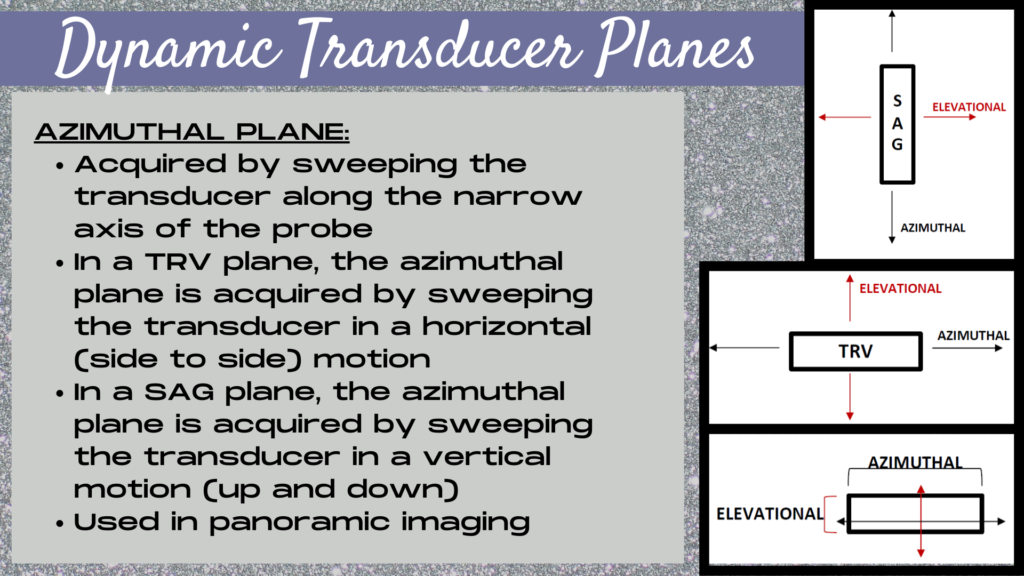
Turn panoramic imaging on, sweep the transducer in an azimuthal direction along the narrow axis of the probe over the area of interest in the desired transducer plane, then hit freeze. This process can be repeated in the orthogonal (perpendicular) transducer plane. What is an azimuthal direction you ask? Azimuthal is a dynamic transducer plane, which explains the motion of a transducer, while a transducer orientation (SAG, TRV, etc.) describes the slice of information that’s being taken through the body. Azimuthal is the direction of motion where the transducer is swept along the narrow side of the probe in order to aquire the frames for a panoramic image.
Other Names For This Control
Various names for this Ultrasound control among different manufacturers include: panoramic imaging, siescape.Conclusion
The key to the perfect Ultrasound image is not getting lucky, or having a thin patient. The real secret to obtaining the perfect Ultrasound is great knoboloby. Your image can only be as good as what you put into it. In this 4 part series we explored 20 essential Ultrasound controls, including Doppler, compound imaging, SRI, harmonics, FOV width and several others, that when applied strategically can make your image shine. Knowledge of these Ultrasound controls only really comes when we are deep in the trenches, struggling to take an image during a difficult to image exam. That’s when we truly understand the essence or spirit of these controls and how a Sonographer is not just a picture taker, but a story weaver. Each image tells a story. And one bad image, or one missing image leaves a hole in our study that affects the story as a whole. Be great! Use these controls to become a master story teller.Check out the other articles in this series: Essential Ultrasound Controls Every Sonographer Should Know And How To Use Them: Part 1; Part 2; Part 3.

